Toronto- July 2024- An international consortium of researchers led by the University Health Network (UHN) in Toronto and the University of Zurich have built the first-ever molecular atlas of the human brain vasculature at single-cell resolution, spanning from early development to adulthood and through disease stages such as brain tumours and brain vascular malformations.
The international consortium includes research teams from UHN’s Krembil Brain Institute, Donald K. Johnson Eye Institute, Toronto General Hospital Research Institute and Princess Margaret Cancer Centre, the University of Toronto’s Donnelly Centre, Mount Sinai Hospital’s Lunenfeld-Tanenbaum Research Institute, University of Zurich, University Hospital Zurich, ETH Zurich, University of Geneva, University Hospital Geneva, as well as collaborators at Weill Cornell Medicine and Memorial Sloan Kettering Cancer Center, in New York.
Researchers have isolated blood vessels from human early developing brains, adult brains, brain tumours and brain vascular malformations. They found that endothelial cells, which line the blood vessels and regulate interactions between the bloodstream and surrounding tissues, behave differently across various stages of brain development, and may have a more important role than previously understood within the brain’s neurovascular signaling networks.
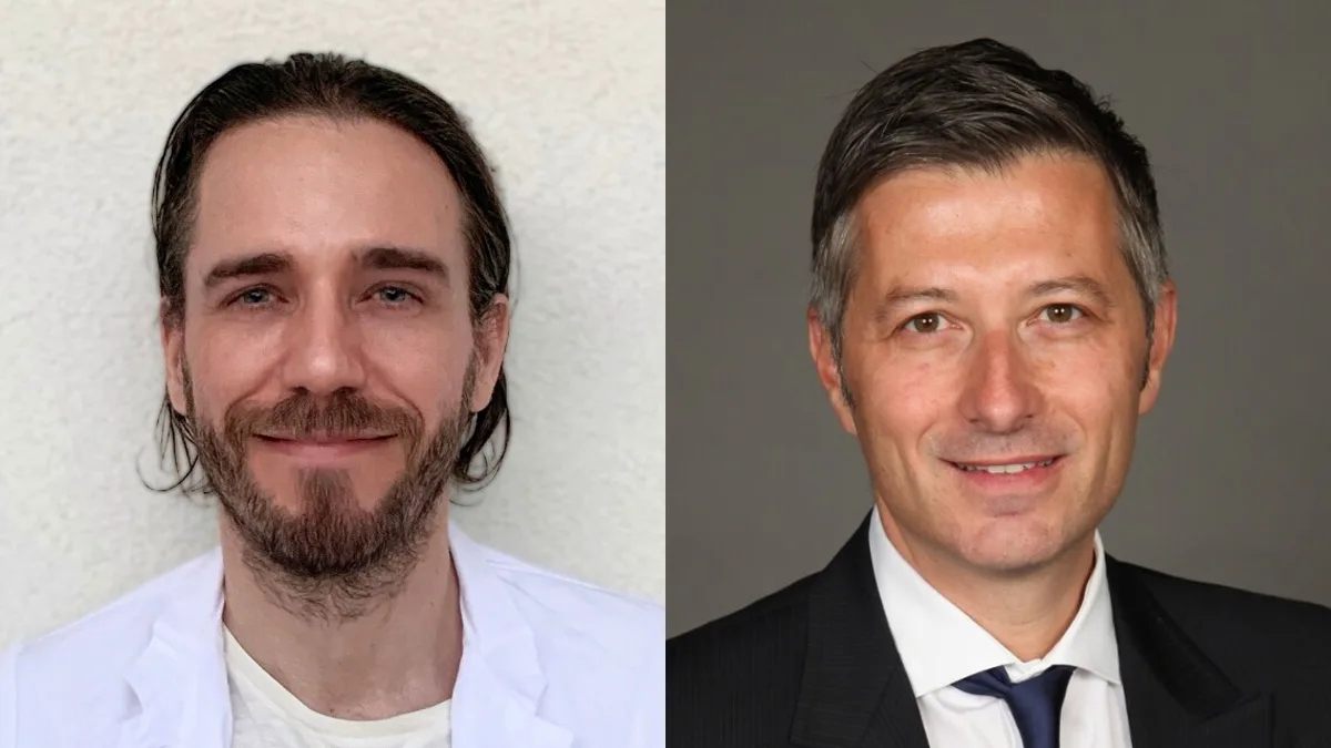
This novel research is published on July 10, 2024 in Nature. “The brain’s vasculature, or blood vessel cells, genes, and pathways, is important for the proper functioning of the healthy early developing and adult brain, as well as for a variety of brain diseases, such as brain tumours, stroke and brain vascular malformations,” says Dr. Thomas Wälchli, corresponding author of the study, and a scientific associate at UHN’s Krembil Brain Institute, a consultant neurosurgeon at University College London (UCL)’s Victor Horsley Department of Neurosurgery, and an Associate Professor/Principal Clinical Research Fellow at the UCL Cancer Institute. “By understanding how these pathways grow and behave during early brain development, how they are silenced in the adult healthy brain, and how they get reactivated in disease, it will provide more insight into the normal functioning of the human brain vasculature and open doors to future therapeutic options.”
Also Read: Latest: Study Reveals How Brain Networks Derive Meaning from Words
“UHN’s Krembil Brain Institute is a high-volume neurosurgical centre, which provided unparalleled access to a clinically diverse patient population. We were able to perform single-cell RNA sequencing of more than 600,000 isolated endothelial, perivascular and other tissue-derived cells from 117 samples, at an unprecedented resolution, giving us an extraordinary look into the inner workings of the brain’s vasculature,” says Dr. Ivan Radovanovic, staff neurosurgeon and Senior Scientist at UHN’s Krembil Brain Institute, Associate Professor at University of Toronto’s Temerty Faculty of Medicine, and co-author of the paper.
“This provides a very large data set that will be an important resource for researchers across the world. By uncovering key differences between healthy and diseased brain vasculature, our work can help identify vulnerabilities of abnormal brain vessels that can be utilized to treat both brain tumours and brain vascular malformations.”


